Some might consider these gross
Atresia Ani Type 2This puppy had NO opening of his colon on the outside of his body. You can see the gas in the x ray coming to a dead end and not out the pelvis, like it should.
It made it to 7 weeks old with never defecating! That's crazy. By the way: he survived and is doing well today! |
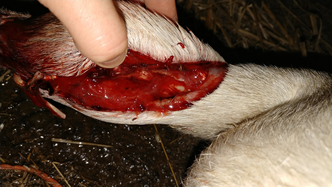
A wrapped wire cut through this sheep's soft tissue leaving the bone exposed.She did well for a repair and is doing great today!
|
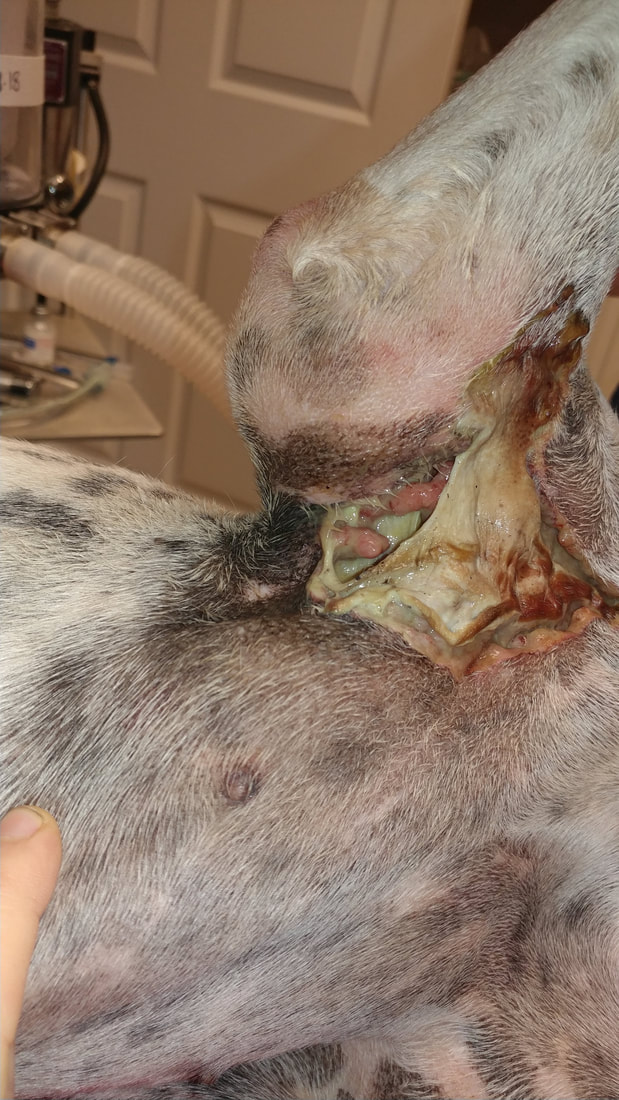
Snake Bite EnvenomationThis is the right armpit of a dog that is known to not give up after one venomous snake bite. She was bitten at least 3 times and it caused her skin to necrose off about two weeks after the bite. She had to have a surgery to make a skin flap to cover this area.
|
|
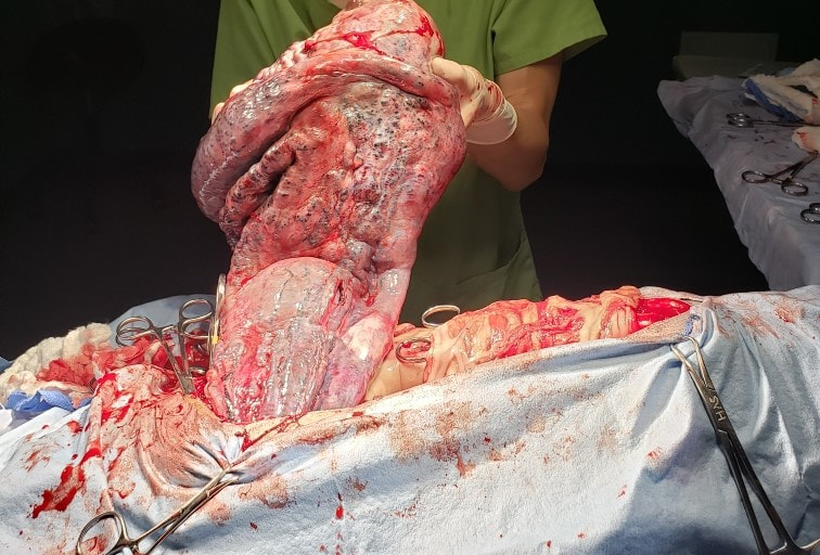
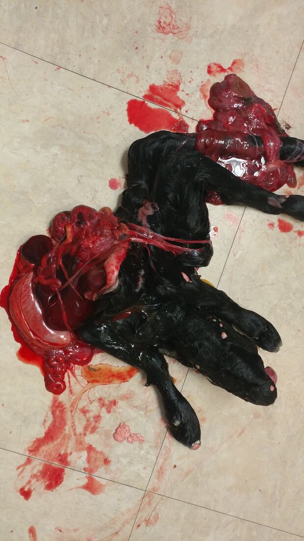
The above picture is hard to tell what is going on, but it is a schistosomus reflexus goat kid. The Mom was having a hard time giving birth and needed a C section. Once we performed the C Section we found out why she couldn't give birth. The insides of the goat kid were growth on the outside and the deformation made it too large for the goat kid to come out of the birthing canal.
|
The back of the skull (top middle) is not smooth like it should be. It had a growth on the bone causing a bump coming up on this dog's head.
The above are two testicles from the same male labrador dog. The testicle on the left is from the UNDESCENDED testicle. So, it was still in his abdomen. The other is from the one that descended appropriately into his scrotum. notice the damage and size difference. (Condition called Cryptorchidism that leads to this)
The below picture is from a testicle from a male dog's abdomen that never was neutered. The internal body temperatures damage a dog's testicle and cause a 7 times likelihood of testicular cancer. That is what this dog below had (DrP in background for reference). This dog's testicular cancer metastasized to the kidneys and he had to be put down. (Condition called Cryptorchidism that leads to this) |


The picture to the right is a foreign body in a canine patient. You can see the "line of demarcation" where the blood supply was compromised as the foreign material in the intestines was causing a blockage. The healthier intestines is at the top of the picture. The compromised tissue is just below this and you can see where it turns dark.
This patient did very well with surgery!
This patient did very well with surgery!

The picture to the left is the left canine tooth on a canine patient. There was severe dental disease present. This shows how the bacteria and disease eats away at the bone around the teeth and there is a hole that extends up into the nasal (nose) cavity. This is called a oronasal fistula. We remove these teeth and have to create a flap to close the large defect.
This doggie is actually super cute and did great!
This doggie is actually super cute and did great!


